Epidemiology of Acute Myocardial Infarction (AMI) and unmet medical needs

What is a heart attack?

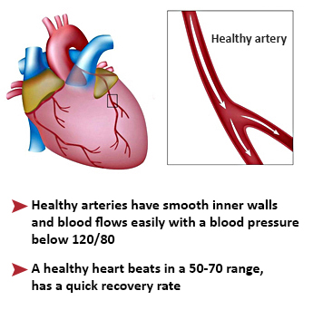
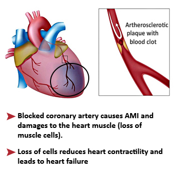
Acute myocardial infarction (AMI), also known as a “heart attack”, is the destruction of an extensive area of the heart muscle. It is mostly the consequence of the obstruction of a coronary artery when cholesterol (fat) accumulates in the form of atheromatous plaques on its walls. A myocardial infarction occurs when an atheromatous plaque breaks off, moves and is trapped in a coronary artery. A blood clot forms around the plaque and interrupts the blood supply, depriving of oxygen the cardiac downstream area which will die.


In France

An average 80 000 people have a heart attack every year, approximately 12 000 of whom will die:

  • One person in 10 dies within an hour,
  • Then the mortality rate is 15% in the first year. However this rate is progressively decreasing for the past years thanks to the speed and quality of intervention by rescue teams (provided they are called quickly), progress in emergency therapies, and increased interventional cardiology units availability and efficacy. 
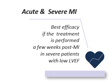
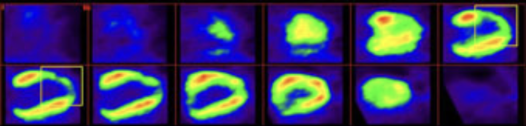
  • However a severe heart attack (about 30% of the cases) leads to a chronic heart failure that weakens heart function and reduces life expectancy (survival rate of 50% at 5 years), despite the daily administration of conventional therapies whose actions are mainly symptomatic.

 Post-AMI heart failure represents a crucial unmet medical need.

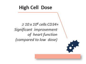
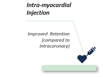
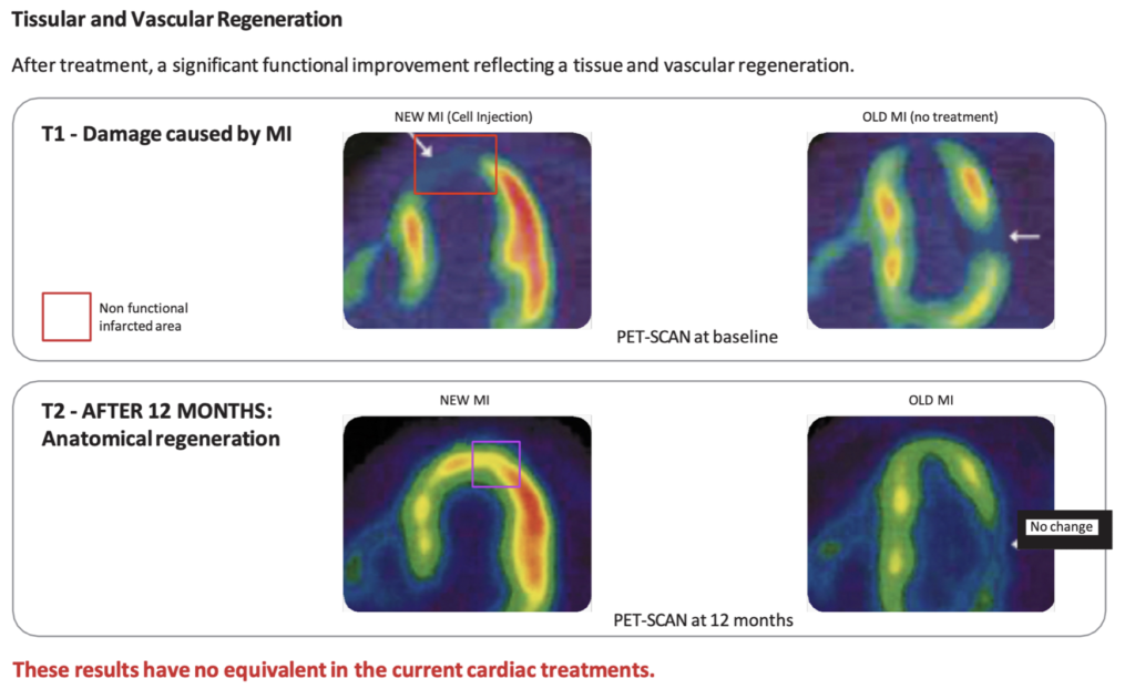
  • Regenerative Medicine thus emerges as a new breakthrough model of medicine that provides a therapeutic solution to cure the disease, being capable to regenerate both structurally and functionally the damaged cardiac area.

Post AMI Cell Therapy- 4 Critical Success Factors

References

1. Hénon P; (2020). Key success factors for regenerative medicine in acquired heart diseases. Stem Cell Reviews and Reports, 16 (3): 441-458

2. Quyyumi, A. A., et al. (2017). PreSERVE AMI. Circulation Research, 120, 324-331.

3. Schächinger, V., et al. (2006). REPAIR-AMI. The New England Journal of Medicine, 355, 1210-1221

4. Mitsutake, Y., et al. (2017). Improvement of local cell delivery using HelixTM transendocardial delivery catheter in a porcine heart. International Heart Journal, 58, 1-645

Successful cell therapy in acute ischemic heart diseases likely depends on four critical factors1:
  • The appropriate cell type: CD34+ stem cells have shown higher potential for tissue regeneration and revascularization compared to other types of cells;
  • The number of cells: Injecting higher CD34+ cell doses has shown good results due to the large numbers of cardiac cells destroyed by AMI;
  • The administration route: Using an intramyocardial injection route via an appropriate catheter is superior to other routes;
  • The timing of injection: Selecting an acute or subacute myocardial ischemia indication, with cells preferentially injected between 4 and 6 weeks after AMI.
These four points appear to be key factors that reduce the likelihood of secondary heart failure, which is very difficult to effectively treat and is associated with a short/ middle term poor prognosis 2,3.
Until now, none of the reported clinical trials have combined all of these key factors.This is the objective of the on-going EXCELLENT trial (EUDRACT 2014–001476-63) using autologous peripheral blood (PB)-CD34+ cells, expanded by the automated StemXpand® device and StemPack® production kits we have developed, and injected trans-endocardially via the Helix™ catheter4 between the end of the fourth and the sixth week post-AMI. CellProthera is the only company who has brought all four factors together.

Pilot study demonstrates potential of CD34+ cell therapy

We had initially shown in a pilot, open label clinical trial that there was a long-term benefit after direct intracardiac delivery of high numbers of autologous PB-CD34+ stem cells in patients with AMI1. For a normal subject, left ventricular ejection fraction (LVEF) is over 70%. The patients included in the study had a LVEF lower than 35%, clearly indicating the presence of a severe heart failure. Three patients were recommended to receive a heart transplant as soon as possible, but they lacked donors. After CD34+ stem cell transplation, LVEF values increased with time, associated with PetScan demonstration of myocardial structure regeneration and revascularization and New York Heart Association grade improvement. Long term follow up of these patients shows that 5 out of 7 patients are still alive with an average follow-up of 17 years (range 16–20 years)2, including all three patients who were initially awaiting a heart transplant but no longer required it after the cell therapy. This is very unusual for patients with such a poor initially prognosis with the additional benefit for them of not needing immunosuppressive treatment as the cell therapy was prepared from their own cells (autologous stem cells). By using adult stem cells obtained from a patient’s own body, this therapeutic approach avoids all challenges currently faced by other types of cell-based therapies, particularly those using allogenic cells (coming from healthy donors), mainly associated with the risk of tissue rejection requiring immunosuppression.

References

1 Pasquet et al. (2009) Long-term benefit of intracardiac delivery of autologous granulocyte–colony-stimulating factor-mobilized blood CD34 + cells containing cardiac progenitors on regional heart structure and function after myocardial infarct. Cytotherapy. 11(8):1002-15.
2 Hénon P et al Transforming Perspectives in Cardiac Cell Therapy: Hypothesis and Commentary Following Updated Results of a Pilot Study Investigating Very Long-Term Clinical Outcomes in Severe AMI Patients Following Trans-Epicardial Injection of Peripheral Blood CD34+ Cells. Stem Cell Rev and Rep 20, 247–257

AMI before stem cells grafting

6 months after stem cells grafting

Myocardial regeneration – Such imaging technique using labeled products like glucose and ammonium allow the quantification of functional recovery (regeneration of myocardial tissue) and also the revascularization of myocardium after cell graft.

EXCELLENT Phase I/IIb clinical trial

JACC: HEART FAILURE Volume 13, Issue 11, November 2025, 102626 | https://doi.org/10.1016/j.jchf.2025.102626

References

1 Roncalli, et al. Transendocardial injection of expanded autologous CD34+ cells after myocardial infarction: Design of the EXCELLENT trial, ESC Heart Failure, 2025 Apr;12(2):1455-1463.

2 Roncalli, et al. Transendocardial Injection of Expanded Autologous CD34+ Cells After Myocardial Infarction: Results of the EXCELLENT Trial. JACC Heart Fail. 2025 Sep 9;13(11):102626.

3 HP Selker, GW Stone et al., Relationship between therapeutic effects on infarct size in acute myocardial infarction and therapeutic effects on 1-year outcomes: A patient-level analysis of randomized clinical trials. Am Heart, 2017, 188:18-25

4 A. Aimo, M. Emdin et al., Prognostic value of reverse remodelling criteria in heart failure with reduced or mid range ejection fraction", ESC Heart Failure, 2021;8(4):3014-3025

5 A. Olivier, P. Rossignol., et al., Combined baseline and one-month changes in big endothelin-1 and brain natriuretic peptide plasma concentrations predict clinical outcomes in patients with left ventricular dysfunction after acute myocardial infarction: Insights from the Eplerenone Post-Acute Myocardial Infarction Heart Failure Efficacy and Survival Study (EPHESUS) study. Int J Cardiol, 2017, 15:241:344-350

The EXCELLENT trial (NCT02669810) was a prospective, multicenter, open-label, randomized controlled study in France and the UK. The objective of the study was to assess the feasibility, safety and efficacy of expanded autologous CD34+ cells (ProtheraCytes®), administered transendocardially to patients with large myocardial infarction (MI) for whom the rate of heart failure hospitalization and death remains high1.

The study enrolled post-MI patients, with an average left ventricular ejection fraction of just 35%, a week after a severe heart attack with significant myocardial damage. Out of the 49 analyzed patients, 33 participants were administered ProtheraCytes® transendocardially on top of standard-of-care, and 16 participants received standard-of-care alone.

Signals of efficacy were observed in the ProtheraCytes® arm in several key markers, predictive of mortality and heart failure worsening:

  • a 9% average reduction in left ventricular end-systolic volume index at 6 months2 (which can predict halving of cardiovascular mortality risk3)
  • a reduction by 50% of severely damaged heart tissue at 6 months (which is linked to a reduced risk of hospitalization for heart failure4)
  • a 70% average drop in NT-proBNP levels at 3 months (which is predictive of improvement in cardiovascular death or hospitalization for worsening heart failure5)

ProtheraCytes® were well tolerated. No patients experienced adverse drug reactions, and there were no deaths. There were nine procedure-related complications in the active group, but all were quickly resolved. Future procedures will be done with an improved injection device and imaging system and rigorous procedure planning.

Over six months, only 1 of the 33 (3%) patients treated required hospitalization for heart failure, compared with 3 of 16 (19%) in the control group, 1 of whom required a heart transplant. In these high-risk patients, CellProthera’s automated expansion platform reliably produced patient’s own expanded CD34+ stem cells at clinical scale. On average, the ProtheraCytes® had a purity over 90% and viability of almost 98%. They were delivered fresh to catheter labs in France and the UK within hours for timely administration.

Patients pathway

When a patient with an AMI is admitted to a cardiology emergency unit, he or she is managed by the medical team. If the patient meets the main initial criteria for inclusion in the clinical trial (LVEF< 50%), the investigating doctor will determine whether the patient is suitable to participate in the study by checking, among other things, the patient’s medical history, current state of health, etc.
If the patient agrees to participate, he/she signs the consent form and participates in the randomization to be enrolled:

  • either in the Verum arm in which the patient will receive the study treatment by injection of autologous CD34+ stem cells, after clinical, biological and radiological (echocardiography, magnetic resonance imagery – MRI – and eventually SpectScan) assessments
  • or in the standard treatment arm, in which the patient receives the standard care commonly performed to treat the condition, and the same assessment programme and follow-up survey as in the Verum arm.

In case the patient is randomized in the Verum arm, he/she will undergo the following steps:

1. CD34+ cell mobilisation
Mobilisation phase of CD34+ stem cells.


2. Enriched stem cells  blood Collection

A blood harvest of 200 ml is carried out on the 6th day.


3. Cell Expansion

The collected blood  is sent to a dedicated cell therapy center and is processed for nine days in our StemXpand® device using single-use StemPack® production kits. This process  allows for the expansion of purified CD34+ stem cells which are then labeled as ProtheraCytes®.

During this phase, the patient is at home.


4. Stem Cell Injection
The day after completion of the expansion process, Protheracytes® are then injected into the patient’s heart by means of a Helix®catheter introduced in the femoral artery and pushed up to the heart’s left ventricle by the interventional cardiologist. The injection does not require a general anaesthesia and takes approximately one hour. It is a one-shot injection, usually performed in the morning, the patient stays one night at the hospital and is discharged the day-after, once the echocardiography control has been done.


Follow-up of the patients (both the Verum and the Control arms)
Control visits at 1, 3 and 6 months are planned for each patient in the clinical trial, comprising clinical and biological evaluation, and echocardiography, MRI, (and eventually SpectScan at 6 months). These visits take place in the hospital where the patients had been initially enrolled in the study. 


Potential benefits

The intracardiac injection of ProtheraCytes® is expected to
  • improve the cardiac ventricular viability and function and therefore improve life expectancy and quality of life
  • reduce the need for pharmacological treatments in case of chronic heart failure
  • serve as an alternative to heart transplant
  • reduce mortality associated with AMI
  • eliminate the risk of graft rejection
This one-shot mini-invasive therapy through the vascular way could soon become an ambulatory procedure which would:
  • lead to significant reduction of overall disease costs for chronic heart failure patients
  • be an ethical technical process using autologous adult stem cells
  • reduce the costs for pharmacological treatments in case of chronic heart failure
  • be  a non-traumatic approach similar to stenting technique
  • reduce costs of existing surgery, transplantation and multiple hospitalisations.